Plankton imaging
Plankton imaging refers to a wide range of techniques and technologies used for quick detection of plankton and its semi-automated recognition. High resolution time series of plankton abundances and biomass are being generated, contributing to knowledge of Belgium’s marine food webs.
Plankton imaging
Plankton imaging yields abundance and biomass information of marine phytoplankton and zooplankton. As it generates a digital copy of a plankter, plankton imaging allows to retroactively go back in time and work on unchanged samples not subject to decay or disintegration. The strength of plankton imaging is seen in the fast processing, allowing for a quick detection of ongoing changes in the marine environment.
Organisms of interest
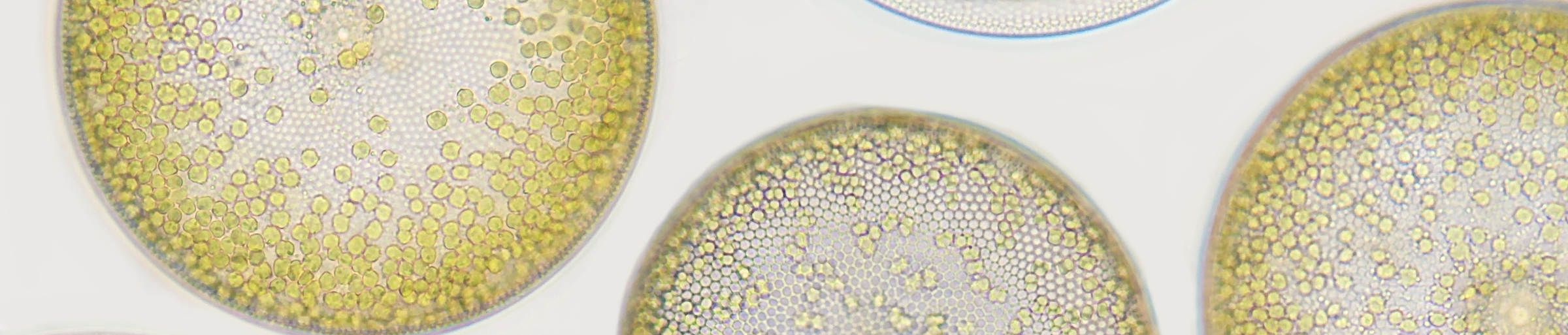
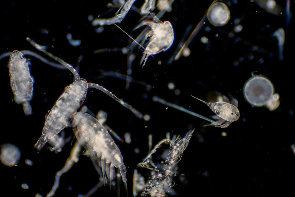
Zooplankton and phytoplankton are two key groups of small aquatic organisms which play important roles in shallow coastal waters. Phytoplankton, consisting of microscopic algae, are primary producers that form the base of the food web in these ecosystems. Zooplankton, a diverse group of small animals, feed on phytoplankton and, in turn, serve as a food source for higher aquatic species. The dynamics of phytoplankton and zooplankton communities are closely linked to the health and functioning of shallow coastal waters and are sensitive to environmental pressures such as climate change, pollution, and habitat degradation.
Infrastructure
Typically, imaging sensors are classified in two groups: in-situ imaging sensors and laboratory-based sensors.
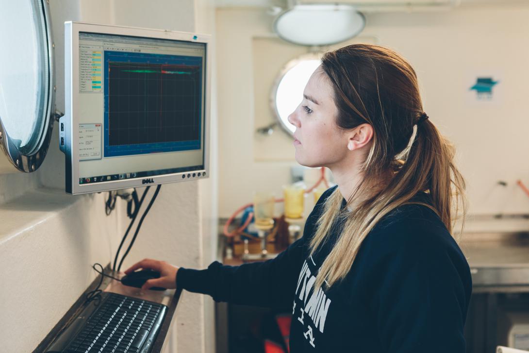
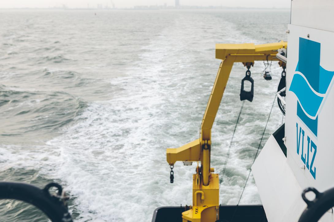
In-situ imaging sensors are deployed in the field by towing them behind a research vessel or by attaching the sensor to a fixed mooring in the water. Three sensors are considered, the Video Plankton Recorder (VPR), the CPICS (Continuous Particle Imaging Classification System) and the Flow Cytometer (FCM). The strength of these techniques is the real-time data collection: particles are photographed in the water in their natural environment. Simultaneous to each picture, field measurements (e.g., seawater temperature, salinity, turbidity, pressure, ...), essential measurements of the depicted particles (e.g. length, height, circularity…) and classification data (e.g., name of the particle that has been photographed) are stored as well. This yields an especially strong dataset important to both marine biologists and geologists.
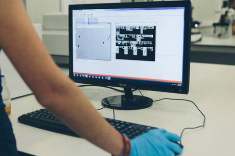
Laboratory-based imaging sensors are deployed in a laboratory and processes collected samples. Two sensors are considered, the ZooSCAN and the FlowCAM (Flow Cytometer and Microscope). Both sensors generate high resolution pictures or vignettes which are analyzed by semi-automatic computer processing: all organisms/particles from the samples are annotated to several taxonomic groups (e.g. Appendicularia, Gammaridae, Calanoida, etc.), but also debris, sand, plastics, etc. The latest machine learning algorithm allows high recognition levels; complementary manual sorting is recommended to achieve a high number of taxonomic groups. Although the resolution of the digitized zooplankton images is lower than that of the images obtained using a binocular microscope, the techniques have proven to be more than adequate for large sample sets.
Spatial coverage
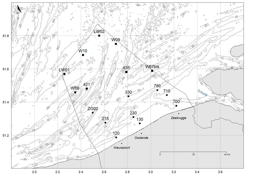
Plankton imaging is a crucial part of the LifeWatch campaigns. During monthly campaigns, zooplankton is sampled at 9 onshore stations; during seasonal campaigns, zooplankton is sampled at 17 on and offshore stations. These samples brough back to OIS and are processed shortly after collecting. All protocols used for plankton imaging are described (Mortelmans et al., 2019; Amadei Martinez et al., 2020) and data is available via the LifeWatch data explorers on zooplankton and phytoplankton. Finally, fixed datasets are released on a yearly basis further contributing to FAIR data, accessible for zooplankton and phytoplankton. LifeWatch further contributes by providing experience to users of the infrastructure.
Data & Services
Related stories
Relevant news
-
SoundLib: The Marine Sound Library for the Belgian Part of the North Sea 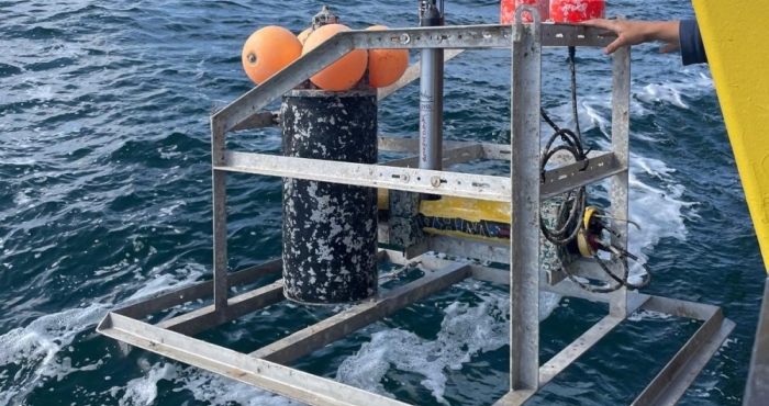
SoundLib: The Marine Sound Library for the Belgian Part of the North Sea
VLIZ has launched the Marine Sound Library (SoundLib), a unique open database of underwater sounds from the North Sea. With thousands of recordings and advanced analysis tools, the platform provides new insights into the region’s acoustic environment. SoundLib supports scientists, policymakers, and the public in understanding how natural and human-made sounds affect marine ecosystems, and creates new opportunities for AI-driven research. -
NELOS divers use WoRMS in digital dive log: a step toward citizen science 
NELOS divers use WoRMS in digital dive log: a step toward citizen science
The Flemish diving federation NELOS vzw (Dutch-speaking League for Underwater Research and Sports) has recently integrated the World Register of Marine Species (WoRMS) into its internal platform DIVES — a Digital Verification System used to log tens of thousands of dives each year. -
EurOBIS celebrates its 20th anniversary with a super-harvest! 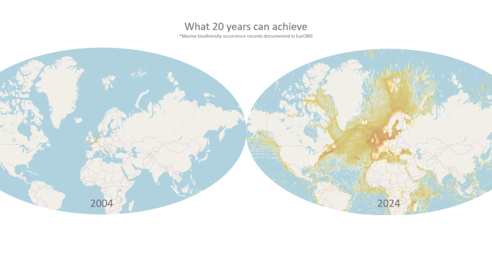
EurOBIS celebrates its 20th anniversary with a super-harvest!
Over its 20 years’ existence, EurOBIS formed alliances with European initiatives as a supporting infrastructure and network. Major milestones include EurOBIS serving as the data backbone of the European Marine Observations and Data Network Biology (EMODnet Biology) since 2009 and being part of the central Species Information Backbone of LifeWatch since 2014. -
Closing the WoRMS 15th anniversary celebrations with an opinion paper 
Closing the WoRMS 15th anniversary celebrations with an opinion paper
In follow-up of the 10th anniversary paper on WoRMS, the Data Management Team is proud to present a paper celebrating the 15th anniversary of WoRMS: "The World Register of Marine Species (WoRMS) through the looking glass: insights from the Data Management Team in light of the crystal anniversary of WoRMS".